Case Summary
A previously healthy young adult presented with a 1-week history of right-sided neck pain, trismus, pleuritic chest pain radiating to the back, shortness of breath, and fever. On admission, the patient was febrile and tachypneic. Initial chest radiography showed multifocal airspace opacities. Laboratory results include leukocytosis (13,900/μL), elevated lactate (2.4 mmol/L), thrombocytopenia (51,000/μL), and respiratory alkalosis (arterial pH 7.46, PaO2 51 mm Hg, PCO2 30 mm Hg).
Imaging Findings
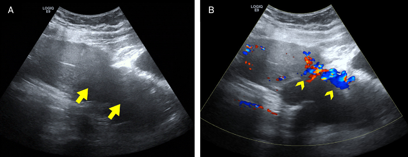
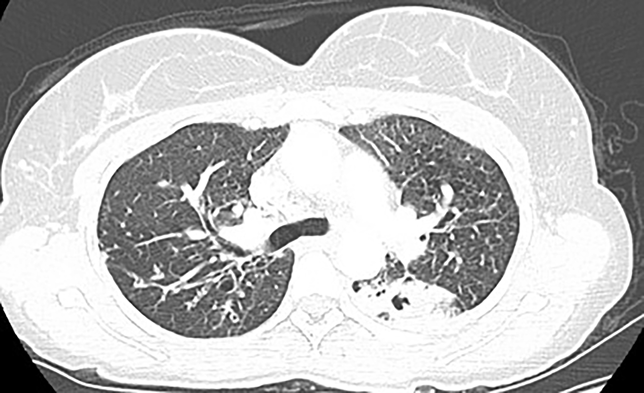
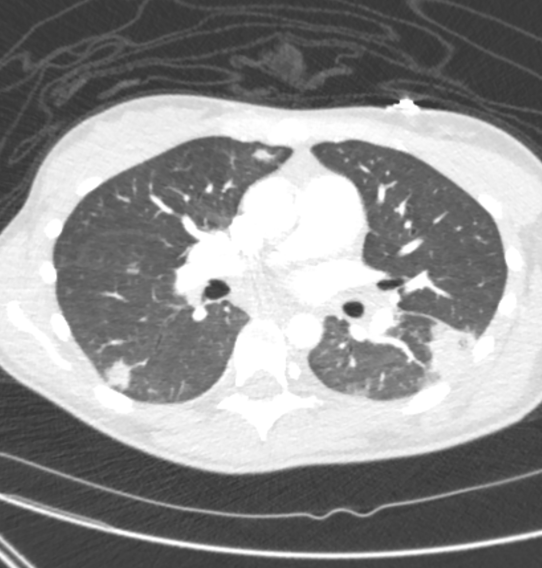
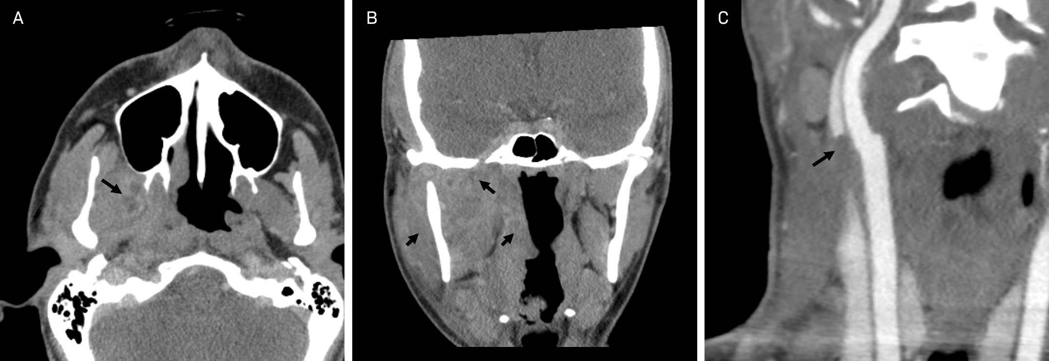
Contrast-enhanced CT of the chest demonstrated multiple bilateral cavitating/consolidative lesions in the lungs consistent with septic pulmonary emboli (Figure 1). Contrast-enhanced CT of the neck demonstrated multispacial infection with phlegmon and inflammatory changes involving the right masticator, pharyngeal mucosal, and parapharyngeal spaces. CTA of the neck confirmed right internal jugular vein (IJV) thrombosis (Figure 2).
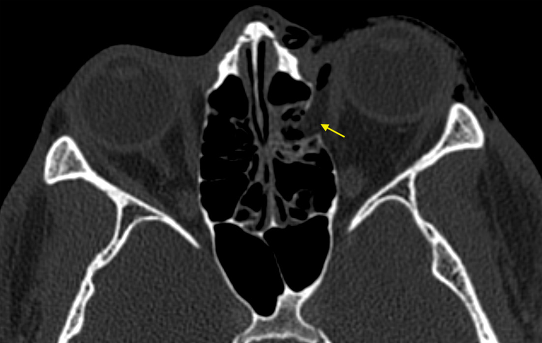
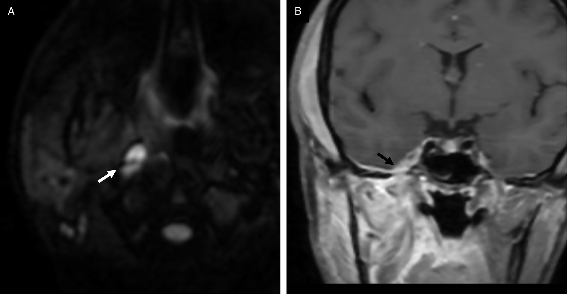
Brain MRI and MR venography demonstrated a small abscess in the right parapharyngeal space and epidural involvement along the right middle cranial fossa with contiguous extension through the foramen ovale (Figure 3), but no cavernous or dural venous sinus thrombosis.
Blood cultures grew Fusobacterium necrophorum.
The patient was treated with intravenous ceftriaxone and metronidazole; their hospital course was complicated by septic shock requiring transient vasopressor support, acute hypoxemic respiratory failure, and acute kidney injury, all of which resolved with supportive medical management. The patient also developed a pleural empyema, which was managed with chest tube placement, resulting in subsequent improvement and resolution. At 1-month follow-up, repeat CT of the maxillofacial region and chest demonstrated resolution of the neck phlegmon and cavitary/consolidative pulmonary lesions.
Diagnosis
Lemierre syndrome with intracranial epidural extension through the foramen ovale
Discussion
Lemierre syndrome is a rare, potentially fatal complication of oropharyngeal infection, classically defined as septic thrombophlebitis of the IJV secondary to F. necrophorum bacteremia. The syndrome primarily affects adolescents and young adults but has been reported across all age groups.1
The disease typically begins with a primary oropharyngeal infection (eg, tonsillitis, peritonsillar abscess, dental infection, or mastoiditis), followed by spread to the parapharyngeal space and subsequent IJV thrombophlebitis. The condition may disseminate through septic embolization, hematogenous spread, or contiguous extension to adjacent structures, including the mediastinum, lungs, or, rarely, the intracranial compartment.2
Intracranial complications (cerebral abscess, meningitis, epidural/subdural empyema, cavernous or dural venous sinus thrombosis) are uncommon; only a few dozen cases in the are documented in the literature compared with many more with pulmonary or systemic septic emboli.1,3-6 Knowledge comes mainly from case reports/series and narrative reviews rather than from large cohort studies.
Intracranial extension of Lemierre syndrome may occur via several routes, including retrograde thrombophlebitis into the dural venous sinuses; septic embolization to brain parenchyma; contiguous extension through skull base foramina; and hematogenous dissemination.3 This case illustrates direct contiguous spread through the foramen ovale, a pathway seldom reported in the literature. The foramen ovale is a skull opening through which the mandibular nerve (cranial nerve V3), lesser petrosal nerve, accessory meningeal artery, and emissary vein exit the skull and enter the masticator space. This creates a direct communication between the intracranial cavity and the masticator space, allowing for the transmission of disease. In our case, the neck infection was centered in the right masticator space extending through the foramen ovale to the intracranial cavity (Figure 5).
Neuroimaging plays a crucial role in detection. Contrast-enhanced MRI and MR venography are the most sensitive modalities for identifying epidural extension, venous thrombosis, or abscess formation. Early imaging should be pursued in any patient with Lemierre syndrome who exhibits neurologic symptoms, severe headache, or cranial nerve deficits.
Management requires prompt intravenous antibiotic therapy targeting anaerobic organisms. Recommended regimens include a beta-lactam/beta-lactamase inhibitor combination or a third-generation cephalosporin plus metronidazole.1 The role of anticoagulation remains controversial and is generally reserved for extensive or propagating thrombosis.7 Previous case reports of Lemierre syndrome with intracranial extension have described multiple brain abscesses and epidural empyema, highlighting the need for central nervous system-penetrating antibiotics and neurosurgical drainage in select patients.4
Conclusion
This case demonstrates a rare instance of Lemierre syndrome with contiguous intracranial extension through the foramen ovale, rather than through the more typical embolic or hematogenous spread. Awareness of atypical routes of spread is important, as delayed recognition may lead to neurological complications. Prompt imaging evaluation and early initiation of broad-spectrum antibiotics remain the cornerstones of management.