Introduction
Soft tissue tumors are a wide class of lesions that occur in nonepithelial, mainly mesenchymal, extraskeletal tissues such as blood vessels, muscles, tendons, fat, peripheral nerves, and fibrous tissue. The incidence of benign tumors is significantly higher than that of malignant tumors. However, rare benign tumors are more common in children.1,2 These tumors are classified according to their genotypic origin and phenotypic variation. Most soft tissue tumors are multifactorial in origin (environmental and genetic factors).3 According to the World Health Organization (WHO 2020) report, soft tissue tumors fall into 11 categories.4 Fibroblastic and fibrohistiocytic tumors are the most common benign soft tissue tumors after fatty neoplasms. Rhabdomyosarcoma, Ewing sarcoma, and synovial sarcoma are common in children, while liposarcoma, leiomyosarcoma, and undifferentiated pleomorphic sarcoma are the most common sarcomas in adults. Soft tissue sarcomas occur most frequently in the extremities (75%), followed by the trunk wall and retroperitoneum.4,5
The use of radiological imaging is of great importance for the evaluation and detection of soft tissue tumors; with the advances made in imaging methods, the presentation of data has increased significantly, which also requires a higher level of expertise to interpret the output data. Therefore, the workload of radiologists has increased, and, on the other hand, due to the complexity of the analysis, there is a need to update their knowledge; as a result, the creation of intelligent systems and algorithms can be a good helper for automatic analysis of images with higher accuracy and speed, and lead to accurate identification of soft tissue tumors with minimal interpretation errors.6
By analyzing medical data, artificial intelligence (AI) can aid in the understanding and timely diagnosis of disease.7 It can also be used to diagnose the type of cancer and determine the appropriate treatment. In addition, AI can be useful in predicting and preventing side effects, as well as assessing a patient’s response to various treatments.8 AI has also recently gained popularity in medical image analysis. Medical imaging assessment with AI has been approved by the US Food and Drug Administration for radiology.9 However, there is insufficient evidence in the assessment of radiological images of soft tissue tumors, and further research is needed to confirm the evidence.10
Considering the importance of the subject and helping to reduce the workload of radiologists, diagnose soft tissue tumors early, with greater accuracy and speed, and help in the treatment process of patients, the present study aimed to evaluate the diagnostic accuracy of AI in soft tissue tumor evaluation in view of radiological images evaluation.
Method
Search and Study Selection
A comprehensive search was conducted using keywords relevant to the study objective in the international databases PubMed, Scopus, Web of Science, and Embase, covering the period from January 1, 2010, to May 25, 2025. All retrieved articles were imported into EndNote software. The study followed the Preferred Reporting Items for Systematic Reviews and Meta-Analyses guidelines.
The PubMed search strategy included Medical Subject Headings (MeSH) related to soft tissue conditions, such as Soft Tissue Neoplasms, Sarcoma, Soft Tissue Infections, Neoplasms, Connective and Soft Tissue, Therapy, Soft Tissue, and Soft Tissue Injuries. It also incorporated specific subheadings for Soft Tissue Neoplasms, including complications, diagnosis, diagnostic imaging, etiology, prevention and control, radiation therapy, rehabilitation, surgery, and therapy.
These terms were combined with imaging-related MeSH terms, including Radiology, Radiography, Radiation Oncology, and the subheading diagnostic imaging, as well as the subheading diagnosis. The strategy further included Artificial Intelligence and Sensitivity and Specificity as MeSH terms.
Additional diagnostic-related terms, such as Diagnosis and Diagnostic Imaging, were included, along with terms related to accuracy and advanced imaging techniques, including Data Accuracy and Radiotherapy, Image-Guided.
Eligibility Criteria
The inclusion criteria were based on the PICO strategy (Table 1). Only human studies published in English were included. Studies evaluating AI applications in radiology were considered, including both prospective and retrospective designs, as well as randomized controlled trials.
| PICO Strategy | |
|---|---|
| Patient/population (P) | Soft-tissue |
| Intervention (I) | Radiology-based artificial intelligence |
| Comparison (C) | Not defined |
| Outcomes (O) | Diagnostic accuracy, diagnostic imaging, sensitivity, and specificity |
The exclusion criteria included case studies of specific cancers and case reports, as well as studies exploring alternative diagnostic methods outside the scope of this review. Studies with incomplete or atypical data reporting were also excluded. Additionally, review articles, laboratory studies, animal studies, letters to the editor, conference papers, and studies without available full text were excluded.
Data Extraction
A predesigned table was used to extract data from the included studies by two independent, blinded authors. In cases of discrepancies, a third author reviewed and resolved them through discussion. A final summary was then prepared based on the collected data.
The data extraction table included the following variables: study name (first author), year of publication, study design, number of participants, and outcomes.
Quality Assessment
Quality assessment was conducted using the Newcastle-Ottawa Scale (NOS),11 which evaluates studies across three domains: selection, comparability, and outcome. Studies with scores greater than seven on the NOS were considered high quality.
For randomized controlled trials, the risk of bias was assessed using the Cochrane Risk of Bias tool (RoB 2), which is the recommended instrument for evaluating bias in randomized trials included in Cochrane Reviews.
Statistical Analysis
The statistical analysis was performed with Stata/MP.v17 as random effect models. Effect size was calculated with 95% CI and the RMEL method.
Results
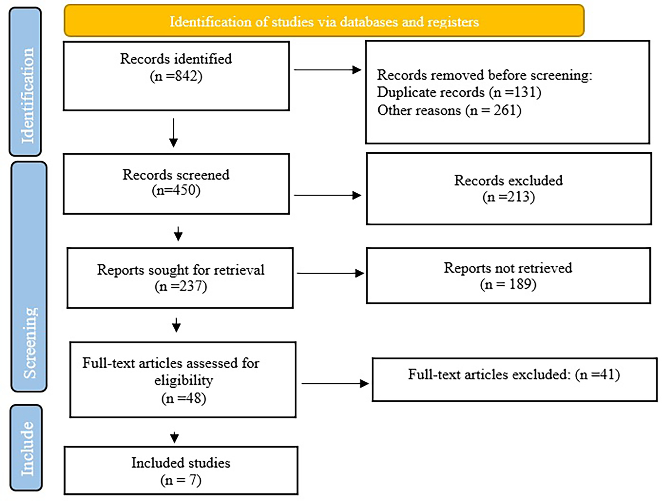
A systematic review of the literature yielded 842 articles that met the search criteria. Based on the exclusion criteria, 392 articles were excluded based on irrelevant or duplicate titles. Abstracts of articles that did not meet the inclusion criteria were removed from a total of 213 screened articles (n = 450). 2 independent, blinded authors reviewed the full texts of 48 articles and screened them for inclusion and exclusion criteria. Only 7 articles met the inclusion criteria and were selected for consideration in this study (Figure 1).
Characteristics of Included Studies
A total of 1230 patients from 7 studies were included in the analysis, comprising 5 retrospective studies and 2 prospective studies. Additional characteristics of the included studies are summarized in Table 2.
| Study | Study Design | Number of Participants | Intervention | Accuracy | Sensitivity | Specificity |
|---|---|---|---|---|---|---|
| Guo et al12 | Retrospective | 185 | Deep learning on MR images | 0.84 | 0.83 | 0.79 |
| Dong et al13 | Prospective | 300 | Machine learning model on endoscopic ultrasonography | 0.91 | 0.9 | 0.93 |
| 1Ye et al14 | Retrospective | 53 | Deep learning on multiparametric MRIs | 0.78 | 0.75 | 0.88 |
| Arthur et al15 | Retrospective | 89 | Machine learning mode on CT | 0.84 | 0.92 | 0.91 |
| Liang et al16 | Retrospective | 126 | Deep learning on MR images | 0.89 | 0.47 | 0.97 |
| Kang et al17 | Prospective | 388 | Machine learning mode on CT | 0.81 | 0.72 | 0.86 |
| Xie et al18 | Retrospective | 89 | Machine learning model, radiographs | 0.68 | 0.57 | 0.91 |
Bias Assessment
Fivestudies received a score of 7-8/9 (low risk of bias) and 2 studies had moderate risk of bias (Table 3).
| Selection | Comparability | Outcome | Total | |||||
|---|---|---|---|---|---|---|---|---|
| Representativeness of Exposed Cohort | Selection of Nonexposed Cohort | Ascertainment of Exposure | Outcome Not Present at the Start of the Study | Assessment Outcome | Length of Follow-Up | |||
| Guo et al12 | * | * | * | * | ** | * | - | 7 |
| Dong et al13 | * | * | * | * | ** | * | - | 7 |
| Ye et al14 | * | * | * | * | ** | * | - | 7 |
| Arthur et al15 | * | * | * | * | ** | * | - | 7 |
| Liang et al16 | * | * | * | * | * | * | - | 6 |
| Kang et al17 | * | * | * | * | * | * | - | 6 |
| Xie et al18 | * | * | ** | * | ** | * | - | 8 |
Diagnostic Accuracy
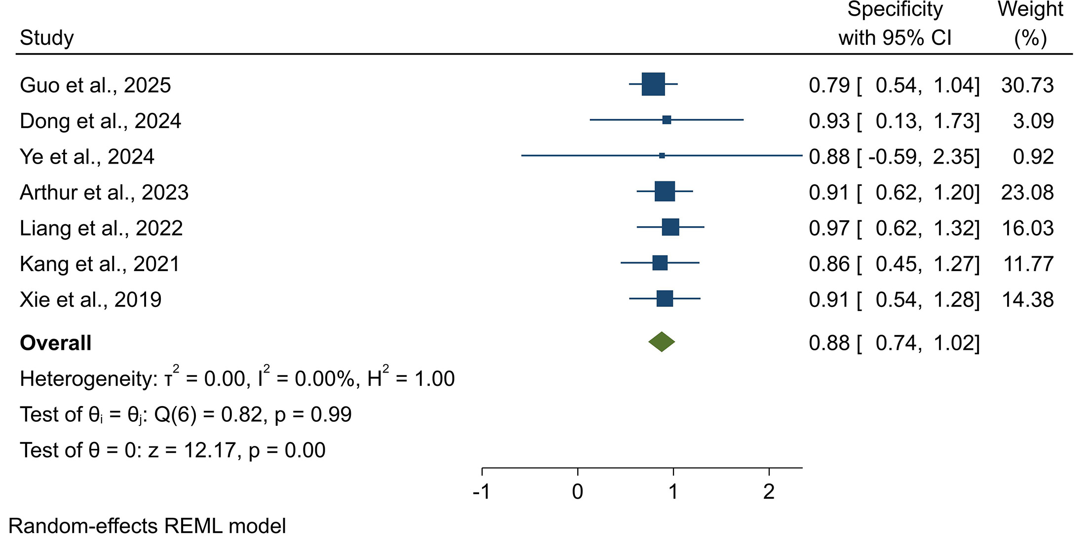
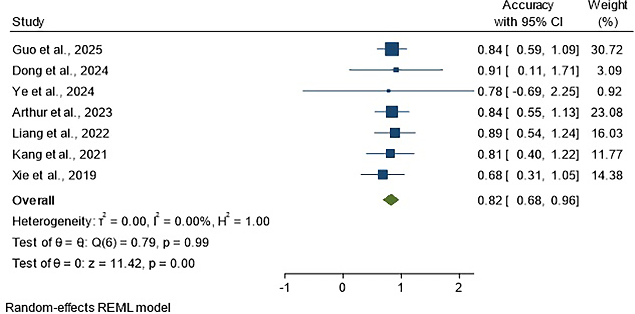
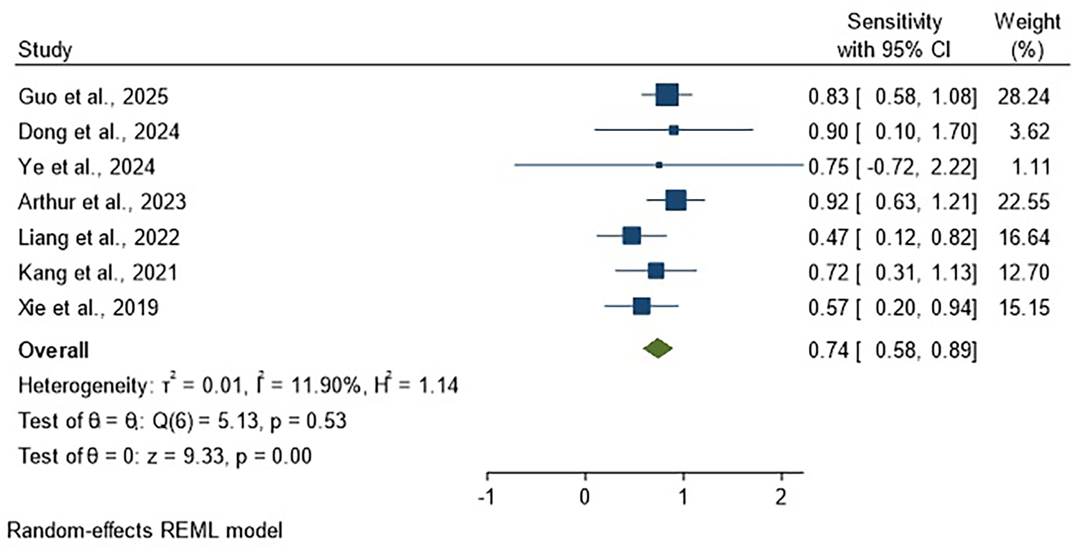
The overall diagnostic accuracy of AI in the assessment of soft tissue tumors based on radiological images was 82% (ES = 0.82; 95% CI 0.68‐0.96) (Figure 2). The sensitivity and specificity were 74% (ES = 0.74; 95% CI 0.58‐0.89) and 88% (ES = 0.88; 95% CI 0.74‐1.02), respectively (Figures 3, 4).
Discussion
Despite advances in research and available evidence, soft tissue tumors remain a significant clinical challenge. Due to their low prevalence, many pathologists and physicians have limited experience with these lesions, which can lead to delays in diagnosis. Late diagnosis is particularly problematic, as advanced or inoperable tumors are more difficult to treat, and overall response rates remain low, ranging from 12% to 24%.19 Therefore, improving diagnostic accuracy and adopting novel methods is of critical importance.
The diagnosis of benign and malignant soft tissue lesions still relies heavily on imaging techniques. The final diagnosis is often achieved through tissue sampling and histopathological interpretation, even though some clinical and imaging features may often help narrow the differential diagnosis. Although malignant tumors are usually larger, a significant percentage of soft tissue malignancies arise from small soft tissue masses. During surgery, these small masses are more likely to be missed or removed less frequently.20 Before tissue sampling, some imaging modalities, primarily contrast-enhanced MRI, may help narrow down the differential considerations, even though histology is still the gold standard for diagnosis. The ability of conventional imaging modalities to accurately distinguish benign and malignant soft tissue tumors, however, has inherent limitations.21
AI has emerged as a tool that can assist radiologists with making timely, accurate interpretations of images, potentially improving patient survival and quality of life.22 According to research, AI can be diagnostically comparable to human interpreting radiologists in a limited number of cases. Due to the complex biomechanical interactions between different anatomical structures, it is difficult for AI researchers to develop reliable algorithms in the face of multiple potential scanning angles and positional changes. Furthermore, complex preprocessing is often required to improve and standardize image quality before AI operations due to field strength, image noise, and acquisition parameter variability.23,24
In the present meta-analysis, AI demonstrated a diagnostic accuracy of 82%, with a sensitivity of 74% and specificity of 88% in the evaluation of soft tissue tumors using radiological images. These findings suggest that AI has considerable potential as a supportive diagnostic tool, enabling faster and reasonably accurate clinical decision-making.
Consistent with the results of the present study, Sudjai et al examining the results of machine learning for distinguishing between ALTs/WDL, observed that AI had high accuracy (88%) in separating muscle lipomas from ALT/WDL, and the performance of this model was comparable to two radiologists with 7-22 years of experience.25 Cay et al reported that AI could predict malignancy in lipomatous masses with a sensitivity of 96.8% and a specificity of 93.72%.26 Fradet et al analyzed MRI results with AI and showed that the AI model could predict malignancy in lipomatous neoplasms with a diagnostic accuracy of 80%,27 while another study found an accuracy of 88% using a radiomics-based nomogram for detecting malignancy in soft tissue masses.28
Heterogeneity among the studies was negligible, indicating that the findings of the present study provide relatively strong evidence. However, most of the included studies were cohort in design, and randomized trials are needed to provide stronger evidence by comparing diagnostic results by AI and radiologists. Despite these limitations, the findings support the integration of AI into radiological workflows as a complementary diagnostic tool.
Although there are encouraging trends in use, there are also a number of barriers and limitations to adoption. Even if AI implementations are validated, achieving high diagnostic accuracy is essential. In order to maximize future applications in daily practice, practicing radiologists should strive to gain a comprehensive understanding of the use cases and challenges associated with implementing AI in prospective clinical workflows. Variability in the way AI applications are implemented is a significant barrier to their use. Neural networks require extensive and often complex training and refinement steps to mimic human cognition. Performance can also be significantly affected by changes in implementation strategies. Standardization supports generalizability and repeatability, increasing diagnostic confidence in potential applications.
Conclusion
Based on the findings of this study, AI demonstrates high diagnostic accuracy, sensitivity, and acceptable specificity in the imaging-based evaluation of soft tissue tumors. AI has the potential to facilitate earlier diagnosis and improve patient prognosis and treatment outcomes. However, the integration of AI into clinical practice remains challenging, and further research is needed to enhance its clinical applicability, address ethical considerations, and optimize its role in radiology workflows.