Pulmonary veno-occlusive disease (PVOD) is an uncommon form of pulmonary hypertension (PH) with preferential involvement of the pulmonary venous system rather than the pulmonary arteries seen in other causes of pulmonary hypertension.1 The condition was initially described by German physician J Hora in 1934; however, not until 1966 was the term PVOD established as a separate entity by Heath, et al.2 In 2015, the Joint Task Force of the European Society of Cardiology (ESC) and the European Respiratory Society (ERS) identified PVOD as a distinct disease, categorizing it as subgroup 1 of pulmonary arterial hypertension (PAH) in the clinical classification of pulmonary hypertension (Table 1).3
Despite the compartmental histological difference, patients with PVOD share clinical characteristics similar to those of patients with PAH due to other causes. However, the distinction of PVOD from other entities of PH is paramount, as patients with PVOD have a worse prognosis.
The gold standard for diagnosis of PVOD is lung biopsy, but this procedure carries a high risk of morbidity/mortality for patients with reduced respiratory reserve.4-6 A consensus for a minimally invasive diagnostic approach has been established which includes diagnostic radiology, pulmonary catherization, echocardiogram, bronchoalveolar lavage, medical therapy, and genetic testing.
This review addresses the various aspects of PVOD, including its clinical presentation, etiology, risk factors, and treatment, and discusses the important role of the radiologist in recognizing key imaging features that can aid in diagnosing the condition.
Clinical Presentation
The clinical presentation of patients with PVOD is nonspecific and difficult to distinguish from PAH and sequelae of cardiovascular disease. Typical symptoms include progressive dyspnea, reduced exercise tolerance, syncope, and hemoptysis.1,4,6-8 Cardiac auscultation may show a prominent P2 component of the second heart sound and systolic murmur of tricuspid regurgitation.4 An additional clinical sign is digital clubbing, which is more frequently seen in PVOD than in idiopathic pulmonary arterial hypertension (IPAH).9 Nonspecific clinical signs include elevation of the jugular venous pressure, peripheral edema, ascites, and hepatomegaly.
Epidemiology, Etiology, and Risk Factors
The incidence of PVOD ranges from 0.1 to 0.5 cases per million.3,10 This estimate is likely low, as 5-10% of cases are either misclassified as IPAH1,5 or unrecognized, as PVOD can occur in association with other diseases, including HIV,12-14 bone marrow transplant, malignancies, and connective tissue diseases.15-21,22,25 Males are usually affected more than females.1,4 The distribution of age at diagnosis is bimodal, with the youngest ranging from 20 to 30 years, reflecting genetic causes, while the second peak is observed at 70 to 80 years, which can be attributed to secondary causes.1
Apart from the mutation in eukaryotic translation initiation factor 2 alpha kinase 4 (EIF2AK4), which has been confirmed as a cause PVOD,26,27 several disease processes and exposures are related to PVOD development. These include inflammatory/immunological, connective tissue diseases, malignancy, medication-related effects, and occupational exposures (Table 2).
| DISEASE ASSOCIATIONS12-14, 29-43 | MEDICATIONS AND EXPOSURES44-48, 28 |
| • Systemic sclerosis | • Radiation |
| • Sickle cell disease | • Mitomycin |
| • Sarcoidosis | • Bleomycin |
| • Pulmonary Langerhans cell histiocytosis | • Cisplatin |
| • Hashimoto thyroiditis | • Vincristine |
| • Hodgkin Lymphoma | • Cyclophosphamide |
| • Primary lung cancer (squamous cell carcinoma) | • Carmustin |
| • Bone marrow transplant | • Oral contraception |
| • Acute myeloid leukemia | • Trichloroethylene |
| • Solid organ transplantation | • Degreasing agents |
| • Human immunodeficiency virus | • Paints |
| • Varnish/glue |
A case control study revealed exposure to trichloroethylene was significantly more frequent in PVOD compared to idiopathic PAH (42% vs 3%, respectively).28 The use of trichloroethylene was reported mostly by metal workers, mechanics, building painters, and cleaners. Other chemical exposures included degreasing agents, paint, and varnish/glue.
The prognosis is bleak for patients with PVOD, with an estimated mortality of 72% at one year from time of diagnosis and close to 100% within two years without definitive treatment as reported in two case series.1,4,49
Histopathology and Pathogenesis
All three vascular compartments of the pulmonary vasculature (pulmonary veins, capillaries and arteries) are affected in PVOD, with a predilection for the pulmonary veins.50,51 Inflammation and immune-mediated injury of pulmonary venules are implicated in PVOD pathogenesis, with the disproportionate upregulation of circulating cytotoxic T-cell, natural killer cell, and natural killer T-cell populations.52
The autosomal recessive bi-allelic mutations of EIF2AK4 have been confirmed in all patients with the heritable form of PVOD and in approximately 9% of sporadic cases.53,54 EIF2AK4 belongs to a family of four kinases that are involved in controlling general translation in response to various cellular stresses. EIF2AK4 function loss leads to increased inflammation and oxidative stress.55
Histologically, diffuse remodeling of post-capillary venules and septal veins results in smooth muscle hyperplasia and intimal fibrosis leading to luminal narrowing. In turn, the affected vessels are obliterated as a result of thrombosis.32,50-52,56 In addition, focal proliferation of capillaries within the alveolar septa is associated with mild interstitial inflammatory infiltrates known as capillary hemangiomatosis-like foci,57 interstitial edema, and/or mild fibrosis of the interlobular septa.2 Pulmonary venous obstruction and pulmonary haemorrhage results in hemosiderin deposition from macrophages. PVOD arterial lesions are similar to PAH; they include intimal fibrosis, medial hypertrophy, and thrombotic occlusion. Plexiform lesions typical of PAH, however, are not present.32,49 Lymphadenopathy secondary to lymphatic vessel congestion occurs frequently in PVOD, leading to nodal congestion with dilatation of the sinuses.58
Imaging Diagnosis
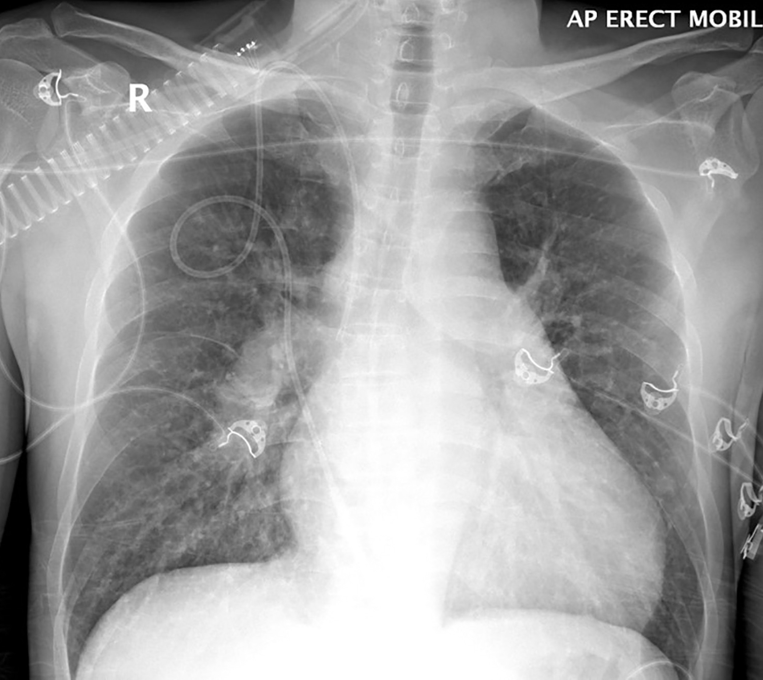
Chest radiography is included in the diagnostic work-up, but the modality has limited sensitivity for PVOD.9,59 In severe pulmonary hypertension, chest radiographs show enlarged main pulmonary arteries and right-ventricular hypertrophy, the detection of which can be aided by a lateral radiograph demonstrating right-ventricular enlargement with encroachment of the right ventricular silhouette into the retrosternal space (Figure 1). Other features include prominent upper-lobe pulmonary vasculature and attenuation of pulmonary vascular markings. Nonspecific findings include Kerley B lines, pulmonary edema, and pleural effusions.6
Thin-section CT, a relatively specific test, plays an important role in the minimally invasive approach to PVOD diagnosis. Interlobular septal thickening and centrilobular ground glass opacities have been shown in several studies to be statistically significant features. The presence of thickened interlobular septa has a sensitivity and specificity of 83% and 87%, respectively, in patients with suspected PVOD.1,62,63,66 Lymphadenopathy is also a common feature (Figures 2, 3).61-64 In the setting of precapillary PAH of unknown etiology, the presence of at least two of three CT signs—septal thickening, centrilobular ground glass opacity, and lymphadenopathy—has a sensitivity of 95.5 % and a specificity of 89 %, with a positive predictive value of 72.5%. A high negative predictive value of 98.5% confers exclusion of PVOD.61
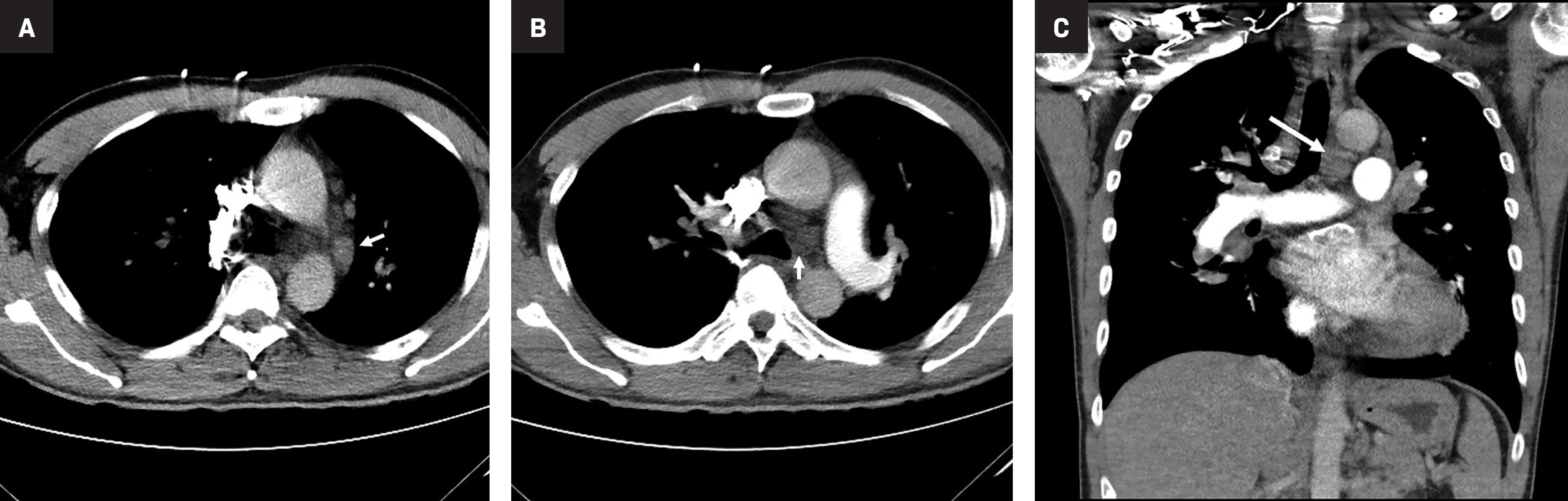
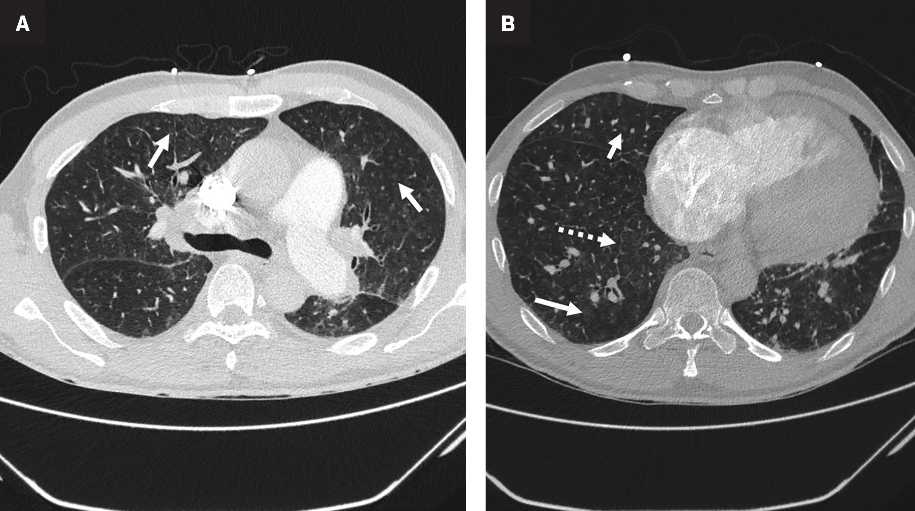
In a small number of biopsies, Resten, et al, found that a centrilobular pattern of distribution of ground glass opacities was significantly more frequent than a panlobular pattern in PVOD, with a sensitivity and specificity of 87% and 67%, respectively.62 The pathogenesis of centrilobular ground glass opacities is not well known; however, their presence may be caused by formation of cholesterol granulomas resulting from occult pulmonary hemorrhage.67 Pleural and pericardial effusion are frequent in PVOD, but they are not specific for pre-capillary PAH.62
Thin-section CT can reveal a mosaic pattern with oligemic areas of hypoattenuation, suggesting vascular rather than small airway disease.65
Diagnostic Criteria
Definitively diagnosing PVOD requires histopathological confirmation via tissue biopsy. This is an invasive procedure, however, that greatly affects patient morbidity and can lead to premature death. A combination of minimally invasive tests has been explored to diagnose PVOD clinically (Table 3). Moreover, Montani, et al, have proposed a PVOD diagnostic algorithm termed “recommended” and “highly probable” PVOD diagnostic criteria (Table 4).28
| TEST | CRITERIA |
| Thin-section CT |
|
| Echocardiogram | Precapillary pulmonary arterial hypertension with elevated tricuspid regurgitation velocity +/- right sided chamber dysfunction32 |
| Right heart catherization | PAWP is usually normal (<15mmHg) mPAP > 25mmHg1,24 |
| Lung function, gas exchange testing | Lower DLCO ABG reveals major resting hypoxaemia1,68 |
| Bronchoalveolar lavage | Haemosiderin-laden macrophages resulting in high Golde scores8 |
Differential Diagnosis
Pulmonary Capillary Hemangiomatosis (PCH)
The European Respiratory Society/European Society of Cardiology (ERS/ESC) Classification of Pulmonary Hypertension has grouped PVOD and PCH in a special subgroup of Group 1 of PAH.3 In the past, PVOD and PCH were regarded as separate conditions. However, current evidence based on histology regards PVOD and PCH as varied expressions of the same disorder,49 while some regard PCH as a secondary process of PVOD occurring in smaller veins.
In addition, there is histological overlap between PVOD and cases of PCH in which they are characterized by aggressive, patch-like capillary angioproliferation.49This is consistent with similar clinical and radiological presentation.
- •
Idiopathic pulmonary arterial hypertension
- -
Absence of interlobular septal thickening
- -
Peripheral lung oligemia
- -
- •
Chronic pulmonary thromboembolism
- -
Absence of interlobular septal thickening
- -
Presence of vascular pruning
- -
PVOD Treatment
The definitive treatment for PVOD is bilateral lung transplantation. This is supported by the 2015 ESC/ERS Guidelines for Pulmonary Hypertension, which recommend listing PVOD patients for transplantation at diagnosis.3,69 Pulmonary veno-occlusive disease was significantly associated with a higher risk of dying while awaiting transplantation compared with PAH.70
A combination of targeted PAH therapy and immunomodulatory and supportive treatments is used to manage PVOD while patients await lung transplantation. Cautious use of PAH drugs such as prostacyclin, endothelin-1, and NO pathway therapies is recommended, as PVOD patients may develop hemodynamic compromise and life-threatening pulmonary edema.71 Approximately 20-50% of PVOD/PCH patients with PAH-specific therapy32,72,73, and 75% of PVOD patients receiving calcium channel blockers experience these complications.32
Treatment with the immunomodulatory drug imatanib as an adjunct to PAH therapy has yielded positive results. Ogawa, et al, reported significant improvement of the World Health Organization functional class and serum brain natriuretic peptide concentration, as well as reduction of mean pulmonary artery pressure and prolonged survival.74
General supportive measures include counseling patients to avoid concomitant medication such as beta-adrenergic receptor blockers, which can precipitate pulmonary hypertension or interfere with the metabolism of vitamin K antagonist anticoagulation therapy, as well as to limit physical activity to tolerance.
Oxygen therapy can be prescribed for individuals with resting or exercise-induced hypoxemia to maintain arterial oxygen saturation greater than 90%.75 Warfarin is routinely used in patients with idiopathic PAH; however, it should be prescribed cautiously in PVOD patients, owing to its association with alveolar hemorrhage.8 Symptomatic right ventricular volume overload may be treated with diuretics under close supervision to avoid hemodynamic compromise.
Conclusion
Pulmonary veno-occlusive disease is a rare entity that causes severe pulmonary hypertension and carries a poor prognosis. Often these patients are misdiagnosed, as PVOD clinical symptoms and signs are similar to those seen in other forms of PAH/PH. Radiologists play an important role in diagnosing PVOD; CT criteria are paramount in the minimally invasive approach to PVOD recognition. Awareness and recognition of centrilobular ground glass opacities, interlobular septal thickening, and mediastinal lymphadenopathy on CT in patients with relevant demographics and risk factors allows for a high index of suspicion for PVOD. This, in turn, can contribute to timely diagnosis and early referral for lung transplantation.