Case Summary
A 68-year-old male presented with a 3-month history of a tender, palpable mass of the left breast in the 11 o’clock position. The patient had a history of prostate cancer, raising concern for metastatic carcinoma.
Imaging Findings
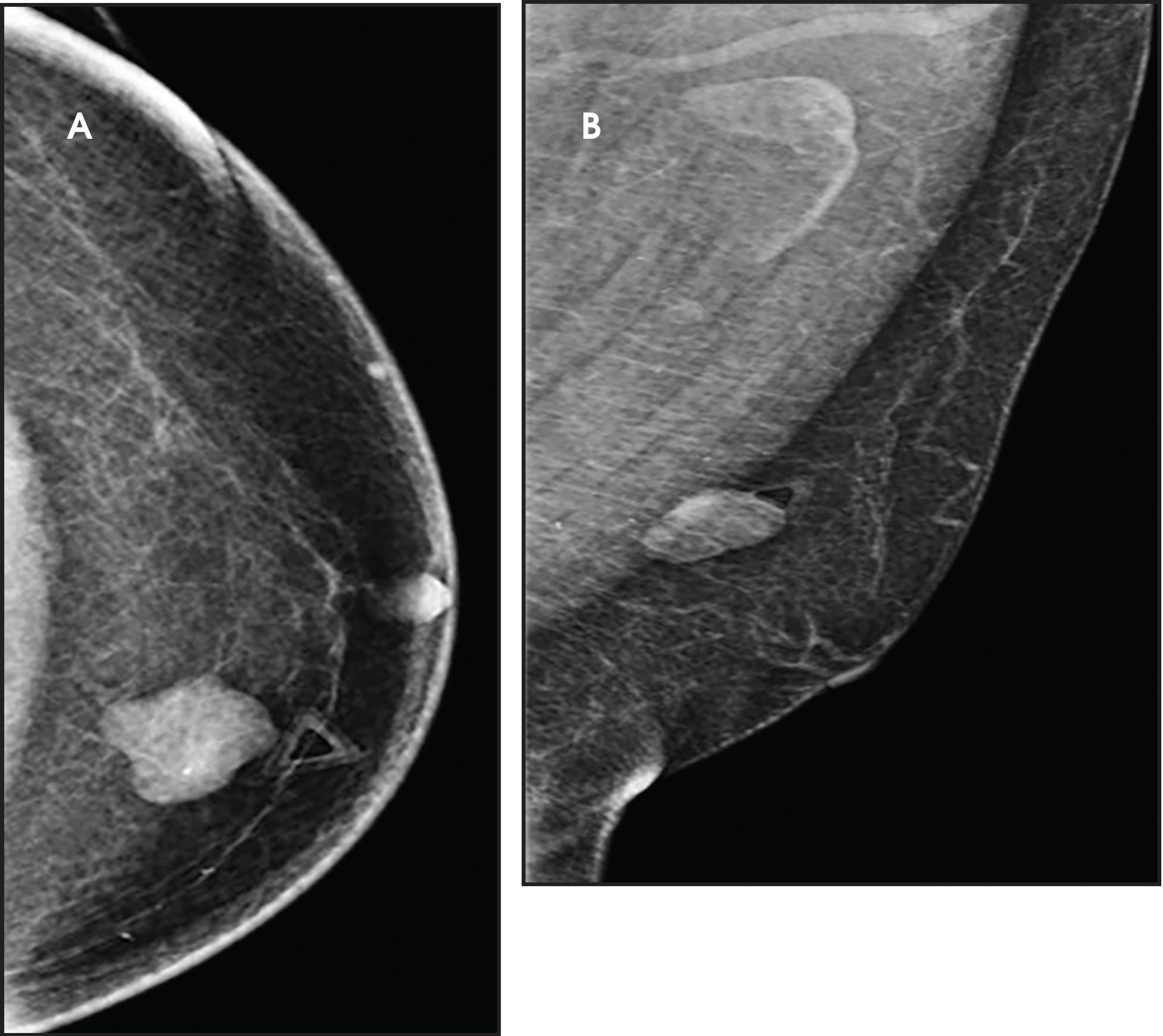
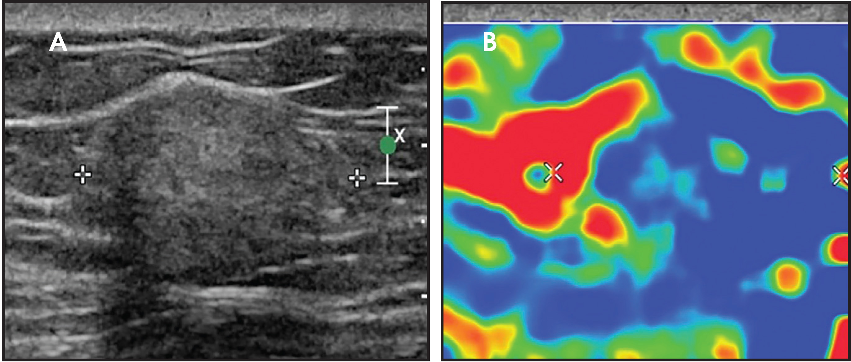
Diagnostic mammography revealed a high-density lesion with macrolobulated margins and without spiculated borders. Scattered calcifications were seen within the left breast. These findings were classified as BI-RADS 4 (suspicious finding). Focused ultrasound demonstrated an elongated hyperechoic mass with smooth margins measuring 2.3 1.2 2.0 cm. There was minimally increased color flow internally. Elastography revealed hard and soft areas within the mass.
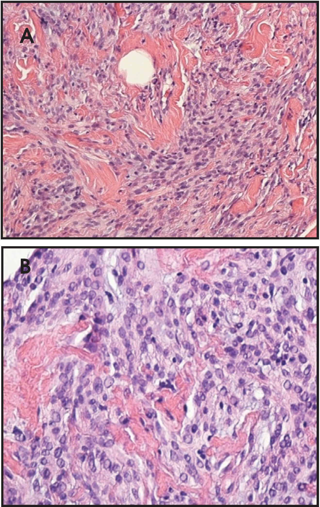
Ultrasound-guided biopsy with a 14-gauge coaxial biopsy gun obtained four core biopsy samples. Pathology results classified the mass as a myofibroblastoma of the breast.
Diagnosis
Mammary myofibroblastoma
Discussion
Myofibroblastomas are uncommon, benign stromal tumors of the breast first described in 1987 by Wargotz, et al.1 Pathologically, myofibroblastomas are described as “benign spindle cell tumors of the mammary stroma”’ due to their derivation from stromal myofibroblasts commonly found in the breast parenchyma.2 Genetically, they are thought to be correlated with the deletion of the 13q14 region, similar to other types of benign mesenchymal and stromal neoplasms.2,3 Myofibroblastomas typically occur in older men and postmenopausal women; however, they can present at any age.4 Although myofibroblastomas are most commonly found within the breast, there have been extramammary cases reported in the literature as well, including in the abdominal wall, the thigh, and even the tongue.3,4,5-8
These masses are typically unilateral, painless, and have the ability to slowly increase in size. The average size is between 1 and 4 cm; however, much larger dimensions have been reported in the literature.9
Mammographically, myofibroblastoma presents as a well-circumscribed mass without architectural distortion or suspicious calcifications.4 They may also demonstrate microlobulated margins that can mimic breast carcinoma on mammography.10 In this case, mammography showed scattered calcifications and a dense mass with macrolobulated margins.
Sonographically, there have been cases where a myofibroblastoma appeared as a circumscribed, slightly echogenic lesion with distal acoustic attenuation, as well as a circumscribed, slightly hypoechoic and non-attenuating lesion.4 Its sonographic appearance may reveal ill-defined borders along with posterior shadowing, and a biopsy is recommended to distinguish it from other lesions.11
Differential considerations include, but are not limited to, primary breast carcinoma, fibroadenoma, gynecomastia and, more importantly in this case, metastatic carcinoma due to the patient’s history of prostate cancer.
Surgical excision is recommended, although ultrasound and/or mammography may be performed every 6 months up to 2 years to confirm stability.12 There have been no reports of malignant transformation; recurrence of myofibroblastoma after surgery is uncommon, likely owing to their smooth margins.
Conclusion
Myofibroblastoma of the breast is a rare, benign mass that is difficult to distinguish from other entities such as a benign fibroadenoma or carcinoma. It is more common in older males and postmenopausal females. Its appearance on mammography and ultrasound is inconclusive, and a core biopsy tissue sample is needed to confirm the diagnosis. Surgical excision is recommended; however, there have been no reports of malignant transformation. Follow-up studies may be performed for up two years to confirm stability.