Case Summary
A middle-aged man presented to his cardiologist with a 1-month history of a newly noticed right breast lump. He had a history of nonischemic cardiomyopathy requiring a heart transplant and had been on immunosuppressants for the past 2 years. His family history was significant for colon cancer in his mother and prostate cancer in his father. He had no personal history of prior malignancy. Given the new palpable breast mass, the patient was referred for dedicated breast imaging for further evaluation.
Imaging Findings
Diagnostic mammography of bilateral breasts was performed with a dermal marker placed over the patient’s palpable lump. In the right retroareolar location, corresponding to the region of concern, a mass with irregular margins was identified with associated nipple retraction (Figure 1). US revealed a correlating 17-mm irregular hypoechoic mass at the right breast 10 o’clock retroareolar position (Figure 2). No abnormal axillary lymphadenopathy was identified on imaging.
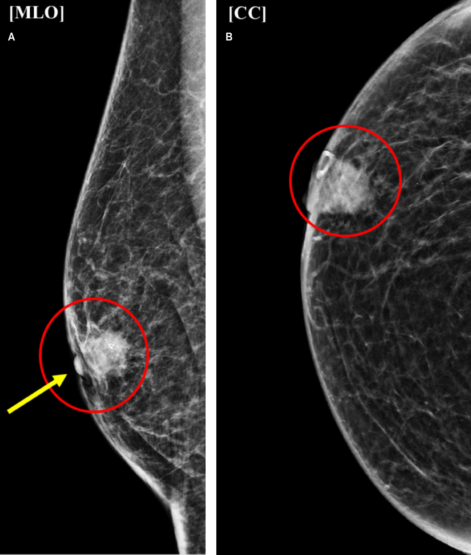
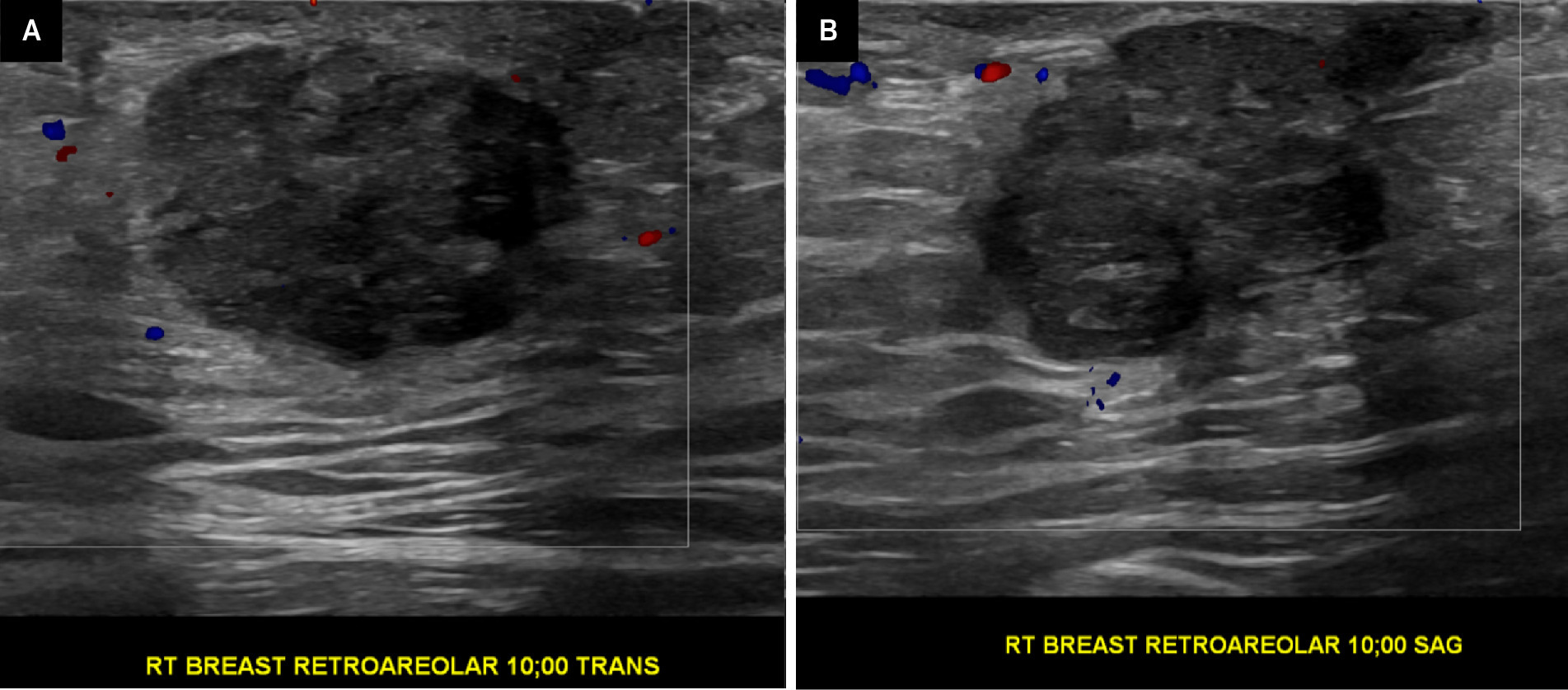
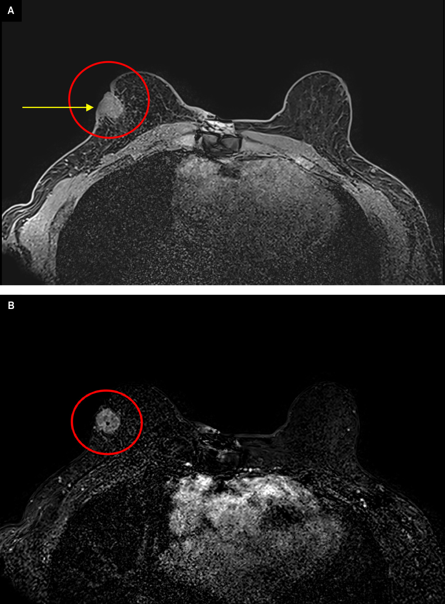
Diagnostic imaging was reported as BI-RADS 4C “high suspicion for malignancy,” with recommendation for US-guided core biopsy. Subsequent US-guided core biopsy demonstrated moderately differentiated invasive ductal carcinoma (IDC) (ER-positive, PR-positive, HER2-negative). Breast MRI demonstrated an 18-mm retroareolar enhancing mass with washout kinetics, associated skin thickening (Figure 3), and nipple retraction.
Diagnosis
Ultrasound-guided core biopsy revealed moderately differentiated IDC (ER-positive, PR-positive, HER2-negative).
The differential diagnosis includes gynecomastia, intraductal papilloma, myofibroblastoma, and metastatic mass.
Discussion
Breast cancer in males is rare, representing less than 1% of the cases of breast cancer. Although genetic factors are commonly discussed in relation to male breast cancer, the cause is largely multifactorial. Occupational exposure to ionizing radiation is associated with an increased lifetime risk of developing breast cancer. Additionally, underlying medical conditions that alter hormonal homeostasis, such as liver dysfunction and thyroid disease, may increase risk. Certain medications can similarly contribute to altering the estrogen/testosterone balance in men, thereby increasing the risk of breast cancer.1 There is no routine screening for male breast cancer, making assessment of risk factors key in detection. The most common presenting symptom in males is a painless, unilateral breast mass with possible secondary symptoms such as nipple retraction and discharge.1
Invasive ductal carcinoma accounts for approximately 85% of male breast cancers. Due to lack of lobular development in males, the risk of developing tumors with a lobular origin, including invasive lobular carcinoma, phyllodes tumors, fibroadenomas, and lobular carcinoma in situ, is less common than in women.2 On imaging, male breast cancer typically presents as a round or irregular mass that is either subareolar or eccentric to the nipple and may demonstrate spiculated or circumscribed margins. Associated findings can include skin thickening and nipple retraction.3 Calcifications may be present but are less common than in female breast cancer. On US, male breast cancers can show varying levels of posterior acoustic enhancement and shadowing as well as varying levels of internal vascularity.2,3 Differentiation of atypical gynecomastia and other benign etiologies may not be possible with imaging alone. US can aid with further evaluation and planning for biopsy when needed.
Despite representing less than 1% of cancer in men, male breast cancer carries a significantly lower 5-year survival rate compared with women.4,5 Age of onset, stage at time of diagnosis, and low index of clinical suspicion are factors that contribute to the male breast cancer mortality rate. In this case, the patient had no history of cancer, negative family history for breast cancer, and unknown BRCA genetic testing, which would indicate a low suspicion for overall risk of breast cancer.3 No formal lifetime-risk assessment is available for males currently as Tyrer-Cuzick and Gail risk assessments are optimized for women.
Additional risk factors include hormonal treatment and certain medications. Estrogen exposure has been linked to an increased incidence of breast cancer in males. In this case, the patient had been taking tacrolimus, an immunosuppressant, for more than 2 years. Chronic immunosuppression is routinely used following solid-organ transplantation and is associated with a higher risk of cancer, most notably nonmelanoma skin cancer.6 Compounding the general risk of malignancy, studies have demonstrated the increased risk of cancer in patients with overall cumulative tacrolimus exposure.7,8 In the absence of traditional risk factors like genetics, this history of organ transplant with chronic immunosuppression may have elevated the patient’s overall risk for cancer. Although the evidence directly linking immunosuppressive therapy and male breast cancer is limited, this case highlights the need for further investigation into a potential relationship between immunosuppression, transplantation, and breast cancer in male transplant recipients.
Conclusion
Male breast cancer is rare, with IDC representing the most common histological subtype. Mortality is generally higher in men than in women, largely due to delay in diagnosis. Mammography and US are valuable tools for detecting breast cancer, although they are not routinely performed in male patients. Despite the lower incidence of breast cancer in men compared with women, it is crucial to perform risk stratification. This includes assessing genetic predisposition, exploring comorbidities and medication history, and performing thorough physical exams. In this case, the patient’s self-examination led to swift dedicated imaging and multidisciplinary treatment evaluations. Although there is limited literature regarding the association between breast cancer and common immunosuppressant therapies such as tacrolimus, being aware of all these potential contributing factors is important and can reduce overall mortality in men through early detection and treatment.